|
|
Ureteritis Cystica
General Considerations
- Rare and benign condition of the ureter and sometimes renal pelvis (pyelitis cystica) resulting from multiple fluid-containing submucosal cysts
- Associated with chronic inflammation and diabetics with chronic and recurring urinary tract infections
- Also associated with nephrolithiasis and chronic obstruction
- Cysts result from degeneration of epithelial cells
- Most often found at 50-60 years of age
- Bilateral in up to 50% of cases
Clinical Findings
- Usually asymptomatic
- May have symptoms of a urinary tract infection
Imaging Findings
- Multiple 2-5 mm filling defects in either the column of contrast rom a retrograde pyelogram or antegrade via CT urography or now, less commonly, an intravenous pyelogram (IVP)
- Rounded lucencies in the contrast-filled lumen which produce scalloping of the margins of the ureter when viewed in profile
- More common in proximal ureter
- Cystitis cystica is almost always diagnosed by cystoscopy
Differential Diagnosis
- Transitional cell carcinoma – usually not as numerous as cysts
- Multiple stones – not as numerous
- Air bubbles – appear round rather than half-moon shaped as cysts do
- Submucosal hemorrhage- extremely rare
Treatment
- The underlying cause of the inflammation or obstruction may be treated
Complications
- Obstruction caused by the cysts has been reported
- Not pre-malignant
 
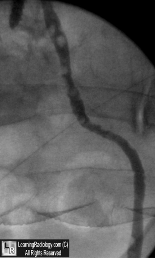
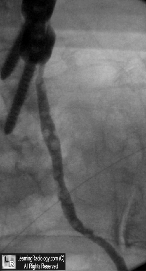
Ureteritis Cystica. Retrograde pyelogram of the left ureter shows multiple,
sharply marginated, rounded filling defects in the ureter, some scalloping the edge
where seen in profile (white arrows) characteristic or ureteritis cystica.
For these same photos without the arrows, click here and here
For more information, click on the link if you see this icon 
Ureteritis Cystica and Pyelitis Cystica: A Review of Cases and Roentgenologic Criteria. BS. Loitman and H Chiat. March 1957 Radiology, 68, 345-351
Ureteritis Cystica: A Radiologic Pathologic Correlation. JG Rothschild and Guan Wu. J Clin Imaging Sci 2011, 1:2
|
|
|
